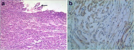
Figure 2.

Histological and immunohistochemical findings of MTSC. (a): the tumor cells are arranged in tubules accompanying spindle cell epithelial components and extracellular mucinous/myxoid matrix were significant; foci papillary structure was seen in case 1 (arrow). (b): Tumor cells were positive for NSE in cases of MTSC.
