
Figure 3.
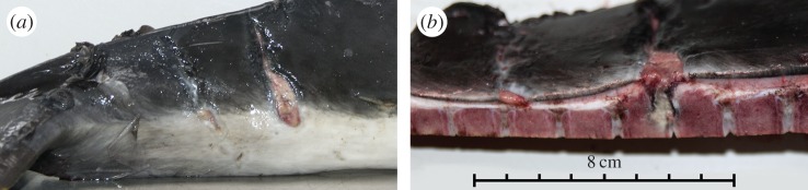
Example of a ‘possible escape’ case. Macroscopic photograph of an inflamed ‘tailstock mark’: (a) lateral view showing a skin wound similar in shape, location and size to ‘tailstock mark’ as shown in figure 1d,f, which shows partial healing; (b) cut section through the tailstock showing the same skin wound and inflammation extending into underlying tissue, new bone formation of the vertebrae and inflammation in the intervertebral disc.