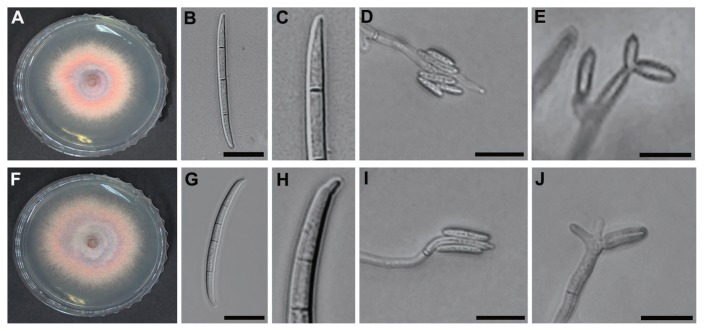
Fig. 3.
Morphological characteristics of F. subglutinans and F. temperatum. (A–E) F. subglutinans and (F–J) F. temperatum. (A and F) Mycelial growth on PDA and (B and G) three-septate (F. subglutinans) and four-septate (F. temperatum) macroconidia on CLA, respectively. (C and H) Close-up view of basal cells of F. subglutinans and F. temperatum, respectively. (D and I) Microconidia in false heads on CLA and (E and J) polyphialides on CLA. Scale bars: 20 μm.

