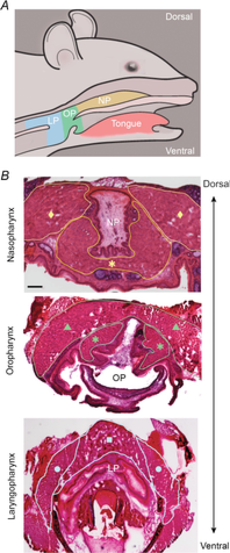
Figure 1. Pharyngeal muscles of mice.

A, murine pharyngeal regions depicting the NP in yellow, OP in green, and LP in blue. B, representative histologic sections of murine pharyngeal tissue stained with haematoxylin and eosin. Pharyngeal muscles are outlined for identification. Representative images of the NP containing the superior pharyngeal constrictor (♦) and palatopharyngeus (*); the OP containing the middle pharyngeal constrictor (▴) and palatopharyngeus (*); and the LP containing the thyropharyngeus (•) and cricopharyngeus (▪) are shown. Bar: 250 μm. LP, laryngopharynx; NP, nasopharynx; OP, oropharynx.
