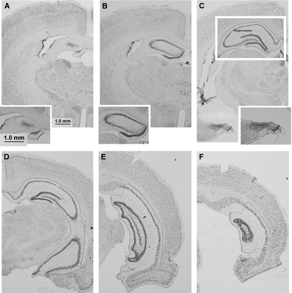
Fig. 2.

Photomicrographs of select coronal Thionin-stained sections through the hippocampus. Dashed lines indicate the boundaries used to define the hippocampus and solid lines those used to define the dentate gyrus. (A,B) Dorsal (septal pole) hippocampus. (C,D) Dorsal and ventral hippocampus. (E,F) Posterior/intermediate hippocampus. Scale bar: 1.0 mm.
