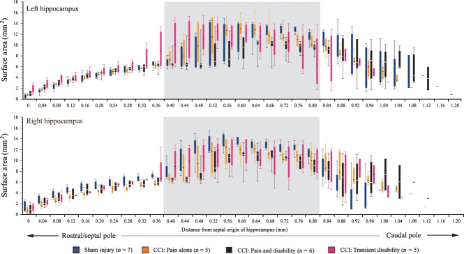
Fig. 5.

Box plots illustrating the means and range of the surface areas of the hippocampus in the following groups: sham-injured control rats (n = 7, blue); Pain alone following CCI (n = 5, yellow); Pain and Disability following CCI (n = 6, black); and Pain and Transient Disability following CCI (n = 5, pink). Serial sections were analysed and plotted from the septal pole (dorsal hippocampus) to the posterior/intermediate pole of the hippocampus. The grey shading highlights the coronal sections where both the dorsal and ventral subregions of the hippocampus are found, which we suggest are not anatomically separable (see text).
