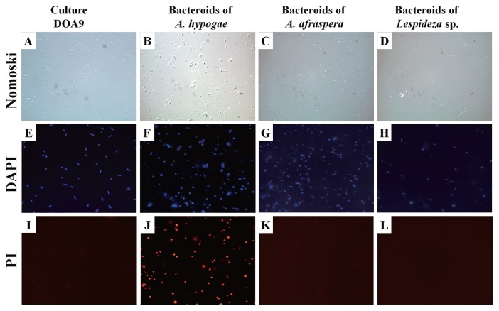
Fig. 4.
Examples of shapes of (left-hand column) free-living DOA9 bacteria and (other columns) resident bacteroids isolated from (B, F, J) Arachis hypogaea, (C, G, K) Aeschynomene afraspera, and (D, H, L) Lespedeza sp. nodules. DIC, differential interference contrast microscopy; DAPI, fluorescence microscopy with 4′,6-diamidino-2-phenylindole stain; PI, fluorescence microscopy with propidium iodide stain.

