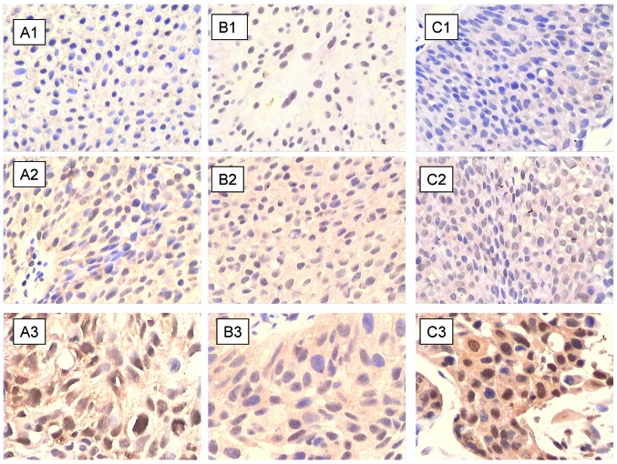
Figure 2.
Immunohistochemical staining of (A) lymphotoxin β receptor (LTβR), (B) phosphorylated (p)-p65, and (C) p52 proteins in different bladder cancer (BCa) pathologic grades: (A1–C1) G1, (A2–C2) G2 and (A3–C3) G3. All three proteins show weak positive staining in G1-, positive staining in G2-, and strong positive staining in G3-grade BCa tissues (×400).

