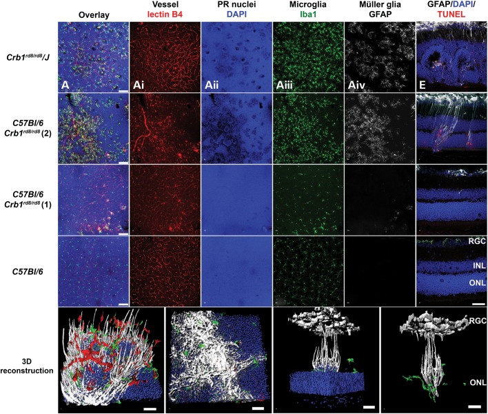
Figure 5.
Localized Müller and microglia activation occurs predominantly in the inferior retina and is associated with dying photoreceptors and vascular changes. (A–D) Overlay of projection images of confocal z-stacks of 30–40 µm height located around the outer plexiform layer (OPL) of the central inferior retina of respective Crb1rd8/rd8 mouse lines and wild-type controls at 2 months of age. For orientation, the deep retinal vascular plexus was labelled by Isolectin B4 (red, Ai, Bi, Ci, Di) and the photoreceptor nuclei of the ONL by DAPI (blue, Aii, Bii, Cii, Dii). Microglia and Müller glia activation in the inner retina was assessed by Iba1 (green, Aiii, Biii, Ciii, Diii) and GFAP (white, Aiv, Biv, Civ, Div) labelling. Scale bar: 75 µm. (E–H) Photoreceptor death in the retina was assessed qualitatively by TUNEL+ labelling (red) on superior to inferior oriented sagittal retinal sections and co-labelled with GFAP (white) for Müller cell activation. Images taken from the inferior central retina are shown. Localized GFAP activation was closely associated with TUNEL+ (red) photoreceptor nuclei (blue). Scale bar: 50 µm. (I–L) 3D reconstruction using Imaris software showing lectinB4+ endothelial cells (red), Iba1+ microglia (green), DAPI-labelled photoreceptor nuclei in the ONL photoreceptor (blue) and GFAP+ Müller cells (white). (I) View at the OPL towards the ONL. (J) Pronounced gliotic scar located at the outer surface of the ONL facing the subretinal space. (J–I) Scale bar: 20 µm. 3D rotated side views of a single inferior lesion with (K) and without (L) representation of the ONL. Columns of photoreceptor nuclei that are elevated from the ONL (K) are surrounded by activated GFAP+ Müller cell processes and Iba1+ microglia (K, L) suggesting a localized glial response around these abnormally arranged photoreceptors. At the retinal ganglion cell layer, GFAP also labels astrocytes next to Müller cell processes. (K, L) Scale bar: 200 µm.

