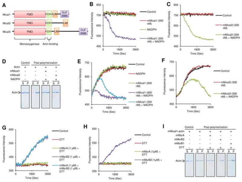
Figure 1.

Mammalian Micals depolymerize F-actin, and MsrBs repolymerize it. (A) Mammals have three Mical genes that code for proteins composed of monooxygenase (FMO), actin-binding CH, LIM, and DUF3585 domains. (B, C) Pre-assembled pyrene-labeled actin (F-actin) was assayed for changes in fluorescence at 407 nm (excitation at 365 nm) in the presence of mMical1 (300 nM), mMical2 (300 nM) and/or NADPH (200 μM). As a control, fluorescence intensity of the pre-assembled pyrene-labeled actin was monitored in the absence of mMical1/NADPH and mMical2/NADPH. (D) Sedimentation/Coomassie staining assay was performed with G-actin before and after polymerization in the presence of mMical1 (1 μM)/NADPH (200 μM) or mMical2 (1 μM)/NADPH (200 μM). S refers to supernatant, and P represents both pellet and the remaining supernatant fraction. Fluorescence at 407 nm (excitation at 365 nm) was monitored for polymerization of pyrene-labeled G-actin incubated with or without (E) mMical1 (300 or 600 nM) and/or NADPH (200 μM) or (F) mMical2 (300 nM) and/or NADPH (200 μM). Actin alone was used for polymerization as a Control in (E) and (F). Then, (G) the mMical1/NADPH- or (H) mMical2/NADPH-treated pyrene-labeled actin monomer was monitored for repolymerization in the presence of mMsrA (1 μM), mMsrB2 (1 μM), and/or DTT (3 mM), or their absence as a control. (I) Sedimentation/Coomassie staining assay was performed with the mMical1 (1 μM)/NADPH (200 μM)-treated actin before and after repolymerization in the presence of mMsrA (1 μM)/DTT (3 mM), mMsrB2 (1 μM)/DTT (3 mM), or mMsrB1-Cys (10 μM)/DTT (3 mM). The mMical1-treated actin alone was used as a control and then this protein was used for further post-polymerization assay. All in vitro biochemical assays used 2.38 μM pyrene-labeled G-actin and all sedimentation/Coomassie staining assays used 4.76 μM G-actin. In the repolymerization assay, pyrene-labeled G-actin (2.38 μM) was first treated with mMicals/NADPH, and the actin monomer was repolymerized following buffer exchange. See also Figure S1.
