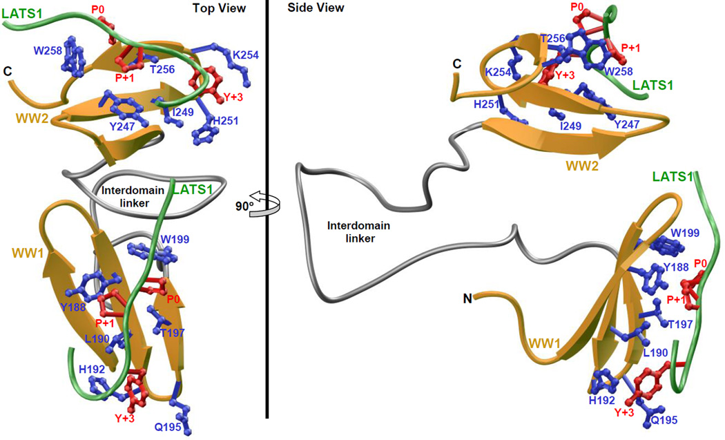
Figure 7.
Structural model of WW1-WW2 tandem module of YAP2 in complex with LATS1 peptide containing the PPXY motif. Two alternative orientations related by a 90°-rotation about the vertical axis are depicted for the inquisitive eye. In each case, the WW domains are shown in yellow with the interdomain linker depicted in gray, and the bound peptide is colored green. The sidechain moieties of residues (blue) within the WW domains engaged in intermolecular contacts with the consensus residues (red) within the PPXY motif of LATS1 peptide are also shown.

