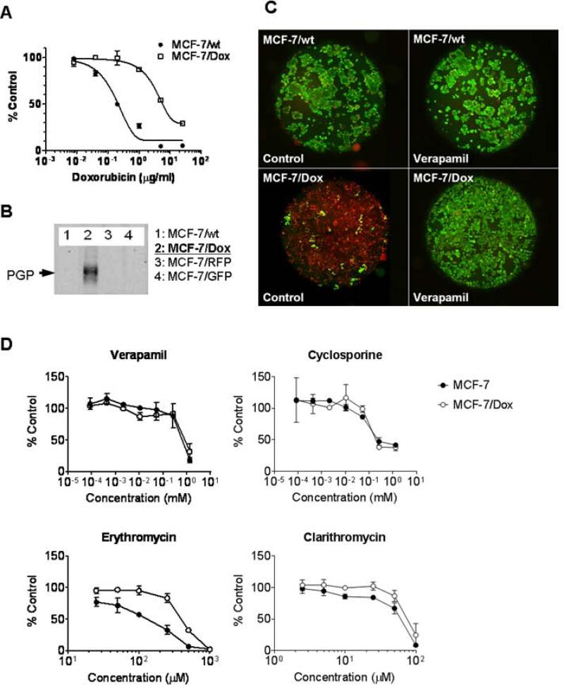
Figure 1.
MDR phenotype upon ABCB1-expression in MCF-7/Dox cell line. A. The survived cell amounts of MCF-7 and MCF-7/Dox after 72 hour culture in the presence of doxorubicin were compared. B. The ABCB1-expression in MCF-7/Dox was confirmed by immunoblot. C. Calcein-AM (green)-exclusion from MCF-7 or MCF-7/Dox in the presence or absence of verapamil (10 μM) was monitored by fluorescence microscopy. Cells were counter-labeled by nuclear staining (red). D. Drug sensitivity to ABCB1-substrates. MCF-7 and MCF-7/Dox were cultured 48 hr in the presence of non-chemotherapeutic agents known as ABCB1 substrates; verapamil, cyclosporine, erythromycin, and clarithromycin.

