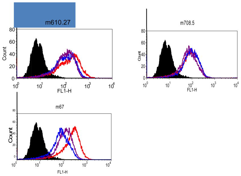
Figure 8. Endocytosis of m67-IGF2 by macrophage-like U937 Cells.
Bound antibodies were detected by FITC-conjugated goat F(ab′)2 anti-human Fc IgG antibody. The black histograms are the blank cells incubated with the secondary antibody only. The histograms for cells incubated with antibody alone, the mixture of antibody and IGF2, and the mixture of the antibody, IGF2 and Cytochalasin D are in red, blue and purple, respectively.

