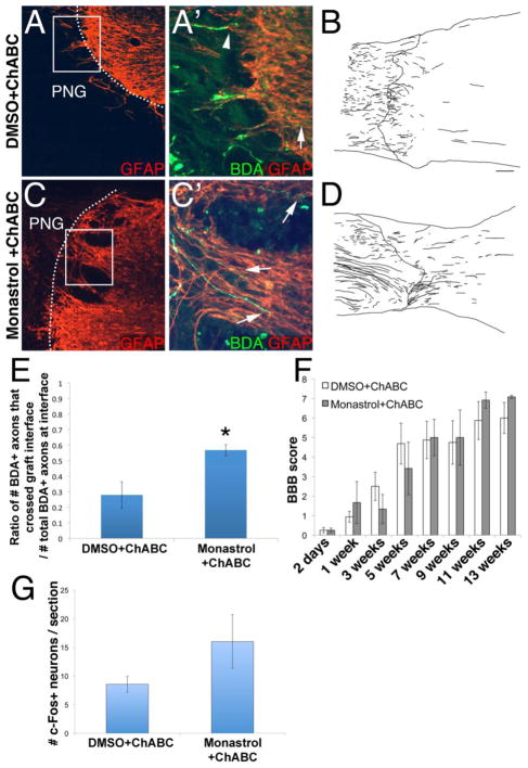
Figure 1. Monastrol treatment enhances regeneration beyond a peripheral nerve graft and through a chondroitinase-treated glial scar.
Axons that regenerated into the graft were labeled with BDA (green). GFAP+ host astrocytes are in red. Insets are high magnification images of boxed regions. While some axons regenerated through the ChABC-treated interface (d), most axons failed to extend into caudal spinal cord (arrows; a′). On the other hand, significantly more axons emerged from the graft (arrowheads, b′, d, e) to reinnervate spinal cord tissue following monastrol treatment. The functional relevance of the axons that regenerated out of the PNG in the ChABC and DMSO or monastrol-treated animals was examined. In the BBB test (f), the scores of both sets of animals improved over time after the injury. However, the scores between the two groups were not significantly different at any of the tested time points. Additionally, spinal cord rostral to the PNG was electrically stimulated. Though this resulted in the induction of c-Fos in neurons caudal to the injury in both groups, there was no significant difference in the number of c-Fos+ neurons (g). c: caudal; scale bar: 100um.; * = p<0.03

