
Fig. 2. AKT elevated ROS in imatinib-treated LPCs but not in LSCs.
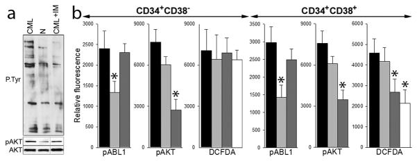
(a) Lin−CD34+cells from normal donor (N) and from CML-CP patient were untreated (CML) or treated with 1 μM imatinib (CML+IM) in the presence of growth factors. Total tyrosine phosphorylated proteins (P-Tyr), AKT phosphorylated on serine 473 (pAKT) and total AKT protein were detected by Western analysis. (b) Lin−CD34+ cells from 3–6 CML-CP patients were untreated (black bars) and treated with 1 μM imatinib (light grey bars), 10 μM perifosine (dark grey bars), or 1 μM imatinib + 10 μM perifosine (white bars) in the presence of growth factors. Phospho-ABL1 (pABL1; phospho-Y245), phospho-AKT (pAKT; phospho-T308) and ROS (DCFDA) were detected in annexin V-negative Lin−CD34+CD38− LSCs and Lin−CD34+CD38+ LPCs as described before 5,8. *p<0.05 in comparison to untreated cells.