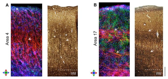
Figure 6.

Comparison of region-specific microstructure of the astriate (primary motor, area 4) and bistriate (primary visual, area 17) cortical areas resolved with dMRI. Track-density maps of area 4 (left panel) and area 17 (right panel) are compared with Bielschowsky silver impregnation of the two areas. Colors in TDI maps denote the local orientation of fibers as indicated by the color indexes. Arrowheads in A indicate a high density of radial fibers resolved in the mid-cortical layers of area 4. Arrows in B mark the distinct bands of tangential fibers resolved in area 17, corresponding to the inner and outer bands of Baillarger. White asterisks mark cortical layer I delineated in dMRI contrasts in both A and B. Scale bar = 0.5 mm.
