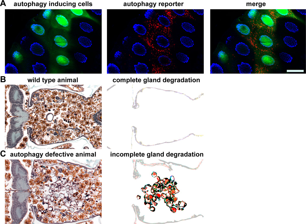
Figure 1. Autophagy in the larval salivary glands of Drosophila melanogaster is necessary for cell death.
A) A larval salivary gland that expresses an autophagy inducing gene only in green fluorescent protein (GFP, green) cells. Autophagy is visualized by using an autophagy reporter that is expressed in all cells and consists of a mCherry tagged Atg8a protein that localizes to autophagosomes and autolysosomes. Nuclei are stained with DAPI (blue). Scale bar represents 50µm.
B) A histology section of a wild type animal in which the salivary glands degrade normally. Note the lack of any salivary gland material (enhanced in the right image). The anterior of the animal is on the left of the image and the posterior of the animal is on the right of the image.
C) A histology section of an animal in which autophagy is defective. Note the large amount of salivary gland material (enhanced in the right image). The anterior of the animal is on the left of the image and the posterior of the animal is on the right of the image.

