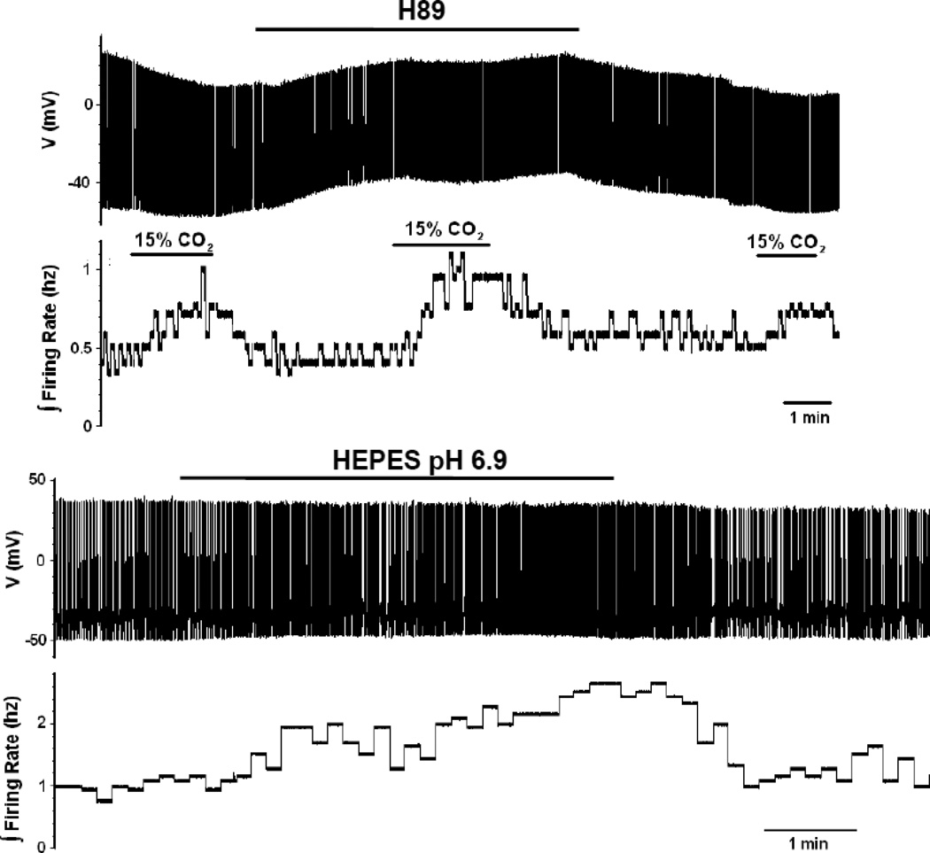
Figure 7.
The effects of PKA inhibition and HEPES-buffered aCSF on the magnitude of the firing rate response of LC neurons from neonatal rats older than P10. (A) The bottom trace shows an integrated firing rate of an LC neuron to 3 challenges of hypercapnia (going from 5% to 15% CO2). Note the modest increase in firing rate in the first and the third challenge, both done in the absence of the PKA-inhibitor H89, but the larger firing rate response in the middle challenge, which is in the presence of H89. The top trace shows a record of individual action potentials at a very slow time trace to show the whole record. (B) The bottom trace shows the integrated firing rate for a neuron that has gone from HEPES-buffered medium at pH 7.45 to an acidified HEPES-buffered medium (6.9). Not the reversible increase in firing rate induced by the acidified HEPES-buffered solution. The top trace shows a record of individual action potentials at a very slow time trace to show the whole record.

