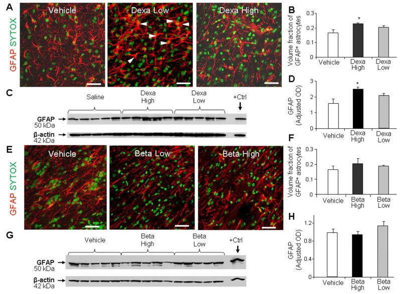
Fig. 3. High-dose dexamethasone induced gliosis, but not the low dose.
A, B) Representative immunofluorescence of cryosections from pup (d 14) treated with dexamethasone labeled with GFAP antibody and sytox (nuclear staining). Note abundant hypertrophic astrocytes in pups treated with high-dose compared to low-dose dexamethasone. Scale bar, 20 μm. Graph shows mean ± SEM (n = 5 each). High-dose, not the low-dose, dexamethasone treatment increased volume fraction of astroglial fibers compared to controls on stereological-analyses. C, D) Western blot analyses for GFAP were performed in forebrain homogenates of pups (14 d). Adult rat brain was positive-control. Graph shows mean ± SEM (n = 5 each). Values were normalized to β-actin. High-dose dexamethasone, not low-dose, increased the GFAP levels compared to vehicle controls. *P < 0.05 for high-dose dexamethasone vs. vehicle controls. E, F) Representative immunolabeling of cryosections from pups (14 d) treated with betamethasone stained with GFAP antibody sytox (nuclear staining). Note astrocyte morphology and density of astroglial fibers are similar in high-dose and low-dose betamethasone treated pups. Scale bar, 20μm. Bar chart shows mean ± SEM (n = 5 each). Neither high-nor low-dose betamethasone treatment affected the volume fraction of astroglial fibers on stereological-analyses. G, H) Western blot analyses for GFAP were performed in the forebrain homogenates. Adult rat brain was positive-control (arrow). Graph shows mean ± SEM (n = 5 each). Betamethasone did not affect GFAP levels.

