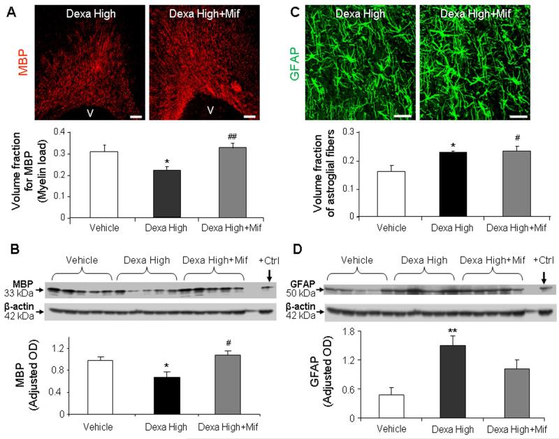
Fig. 4. Dexamethasone-induced hypomyelination was reversed by mifepristone treatment, but not dexamethasone-induced gliosis.
A) Representative immunofluorescence of cryosections from pups (d 14) treated with high-dose dexamethasone and high-dose dexamethasone plus mifepristone, labeled with MBP antibody. Note more abundant immunoreactivity for MBP in pups receiving dexamethasone with mifepristone compared to dexamethasone alone. Scale bar, 100 μm. Graph shows mean ± SEM (n = 5 each). B) Western blot analyses for MBP were performed in the forebrain homogenates of pups as indicated. Adult rat brain was positive-control (arrow). Graph shows mean ± SEM (n = 5 each). MBP levels were less in dexamethasone-treated pups compared with vehicle or dexamethasone plus mifepristone treatment. C) Cryosections from high-dose dexamethasone and high-dose dexamethasone with mifepristone treated pups were immunolabeled with GFAP antibody. Note comparable immunoreactivity for GFAP in dexamethasone, and dexamethasone with mifepristone treated pups. Scale bar, 20μm. Graph shows mean±SEM (n = 5 each). D) Western blot analyses for GFAP were performed in forebrain homogenates of pups. Graph shows mean ± SEM (n = 5 each). GFAP levels were comparable between pups receiving dexamethasone with mifepristone and dexamethasone alone. For Fig. A and B: *P < 0.05 high-dose dexamethasone vs. vehicle. #P < 0.05 high-dose dexamethasone (dexa high) vs. dexamethasone + mifepristone (Mif). For Fig C and D: *P < 0.05 for high-dose dexamethasone vs. vehicle. #P<0.05, for vehicle vs. dexamethasone + mifepristone.

