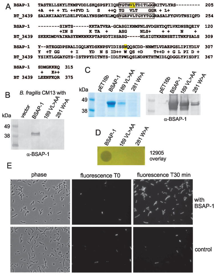
Figure 5.
MACPF domain is necessary for BSAP-1 activity. A. Alignment of the MACPF domains of BSAP-1 and BT_3439 of B. thetaiotaomicron VPI-5482. A conserved MACPF motif is boxed with residues altered by site-directed mutagenesis highlighted in yellow. B. Western blot of supernatants of B. fragilis CM13 containing the gene for WT BSAP-1, or BSAP-1 site-directed mutant genes, probed with antiserum to BSAP-1. C. Coomassie-stained gel (left panel) or western immunoblot (right panel) showing the purification of His-tagged BSAP-1 or BSAP-1 site-directed mutants from BL21/DE3 induced at room temperature. The coomassie-stained gel shows the unconcentrated mutant proteins following purification whereas the western blot shows reactivity when the mutant protein samples were each concentrated 10-fold. The blot was probed with antiserum to BSAP-1. D. Analysis of the killing of strain 12905 in an agar overlay by purified BSAP-1 or concentrated BSAP-1 site-directed mutants E. Microscopic analysis of PI incorporation by strain 12905 exposed to BSAP-1 contained in culture supernatant or control supernatant. Fluorescence images are shown at the starting point and 30 minutes later.

