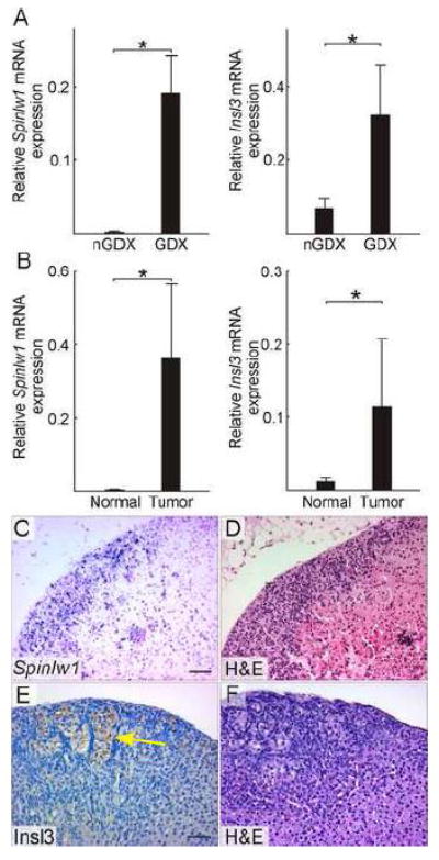
Figure 3. Two genes identified by microarray expression profiling, Spinlw1 and Insl3, are upregulated in GDX-induced adrenocortical neoplasms of the mouse.

(A) qRT-PCR analysis of Spinlw1 and Insl3 in whole adrenal mRNA from non-gonadectomized (nGDX) or gonadectomized (GDX) female DBA/2J mice (n = 4 per group). (B) qRT-PCR analysis of Spinlw1 and Insl3 in normal or tumor tissue isolated by LCM from the adrenal cortex of ovariectomized DBA/2J mice (n = 4 per group). qRT-PCR results were normalized to expression of the housekeeping gene Actb (*, P < 0.05); normalization to Gapdh yielded similar results. Statistical significance was determined using ANOVA with Dunnett’s multiple comparison test. (C) In situ hybridization of adrenal tissue from an ovariectomized DBA/2J mouse using a Spinlw1 antisense riboprobe. (D) H&E staining of an adjacent tissue section. Spinlw1 mRNA localized to neoplastic cells in the subcapsular region. (C) Immunoperoxidase staining of adrenal tissue from an ovariectomized DBA/2J mouse using anti-INSL3. (D) H&E staining of an adjacent tissue section. INSL3 immunoreactivity was evident in lipid-laden type B neoplastic cells [see (Bielinska et al., 2006) for a description of this cell type]. Control experiments demonstrated INSL3 immunoreactivity in Leydig cells of the adult testis (data not shown). Bars = 50 μm.
