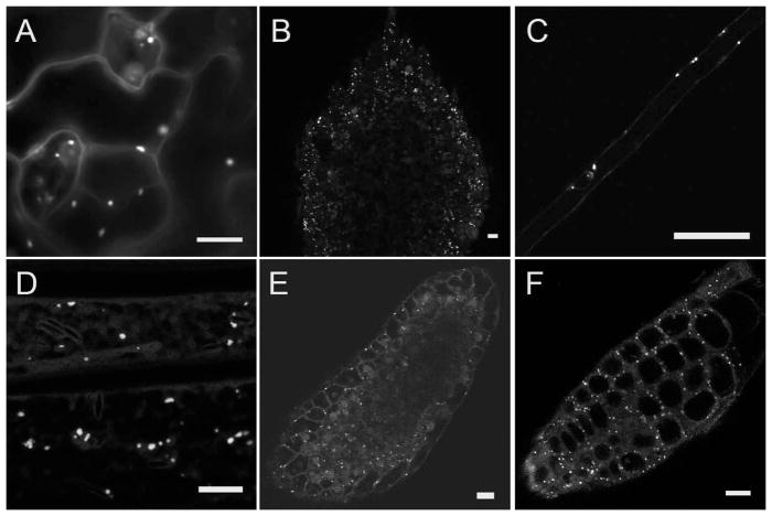
Figure 1.
GFP-cDNA fusion proteins in Arabidopsis lines CS84743 and CS84812 accumulate in circular structures about 1μm in diameter. Live samples of lines CS84743 (A–C) and CS84812 (D–F) were observed by epifluorescence and laser scanning confocal microscopy. Fluorescent structures were observed in all organs examined, including leaf epidermis (A), cotyledons (B), root hairs (C), etiolated hypocotyl epidermis (D), leaf primordia (E), and root tips (F). Faint GFP fluorescence is sometimes visible in nuclei and in the cortical cytoplasm. Scale bars = 10μm.

