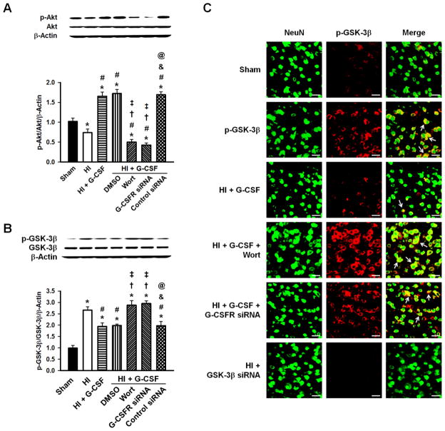
Figure 5.
Representative Western blots and densitometric quantification of p-Akt/Akt/β-Actin and p-GSK-3β/GSK-3β/β-Actin 48hrs after HI. A, p-Akt showed a decrease after HI, however it was significantly elevated after G-CSF was administered which was abolished by ICV injection of G-CSFR siRNA or Wortmannin. No changes were seen in total Akt. B, G-CSF robustly reduced the p-GSK-3β/GSK-3β ratio compared to HI group, which was blocked by Wortmannin and G-CSFR siRNA. DMSO and Control siRNA administration groups were similar to the G-CSF treated group. No changes were seen in total GSK-3β. n=6/group. C, Localization of p-GSK-3β (red) with neuronal marker NeuN (green) was observed. GSK-3β siRNA group showed no expression of GSK-3β. n=3/group. Scale bars=25μm. * p<0.05 vs. Sham, # p<0.05 vs. HI, † p<0.05 vs. HI + G-CSF, ‡ p<0.05 vs. HI + G-CSF + DMSO, & p<0.05 vs. HI + G-CSF + Wortmannin, @ p<0.05 vs. HI + G-CSF + G-CSFR siRNA.

