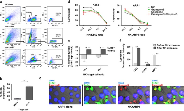
Figure 5.
The mechanisms underlying CB-NK-mediated cytotoxicity differ between target cells: (a) Degranulation (CD107a expression) of CB-NK after 4 h of incubation with K562 or ARP1 cells. (b) Mean±S.E.M. values from a representative experiment shown in a (n=4). (c) GrB expression in ARP1 cells after being co-cultured with CB-NK for 20 min. ARP1 cells were stained in blue (CMAC) and CB-NK in green (bodipy). GrB is indicated in red. (d) CB-NK cytotoxicity reduction after inhibiting GrB and Caspase-3. Killing is shown for control CB-NK, after inhibiting GrB in CB-NK, Caspase-3 in target cells and both, GrB in CB-NK and Caspase-3 in target cells. (e) Percentage of killing reduction after inhibiting GrB and Capase-3 in four different CB units of the representative experiment shown in d. (f) Lysosome levels before and after CB-NK exposure for 40 min for K562 cells, CD19+ cells from a healthy donor and ARP1 cells. Bars represent mean±S.E.M. *P≤0.05. **P≤0.0001

