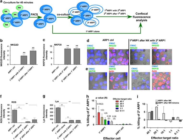
Figure 8.
CB-NK cytotoxicity is transmissible between MM cells: (a) Design experiment for results from b to h. Bodipy-labeled CB-NK and CMAC-labeled ARP1 cells (1° ARP1 cells) were co-cultured in cell pellet for 40 min, then 1° ARP1 cells were FACS sorted and half of them were left alone (1° ARP1 alone), and half of them (1° ARP1 with 2° ARP1) were co-cultured in cell pellet for 40 min with fresh unstained ARP1 cells (2° ARP1 cells after 1° ARP1), then confocal fluorescence experiments and analyses were performed discriminating between the two sets of ARP1 cells based on 1° ARP1 cells labeled in blue (CMAC) and 2° ARP1 cells unstained. (b and c) NKG2D (b) and NKP30 (c) expression in 1° ARP1 cells and 2° ARP1 cells. Bars represent mean±S.E.M. (d and e) Representative images of NKG2D (d) and NKP30 (e) expression in green in both sets of ARP1 cells of values obtained in b and c. Actin is shown in red. Lysosome (f) and ROS (g) expression in the two sets of ARP1 cells. ARP1 ctrl: indicates ARP1 cells alone sorted as control for the experiments. Bars represent mean±S.E.M. Results from b to g were confirmed in three different experiments. (h) Cytotoxicity assay performed adding as effector cells 1° ARP1 sorted cells and as targets fresh ARP1 cells. Effectors were added at different effector:target cell ratios. In parallel, ARP1 cells alone were sorted and added as effectors to the cytotoxicity assay as control. (i) Cytotoxicity reduction of 1° ARP1 (as effectors) versus fresh ARP1 cells (as targets) after adding (ARP1 after NK+stroma) or not adding (ARP1 after NK) stroma to the assay. *P≤0.05. **P≤0.0001

