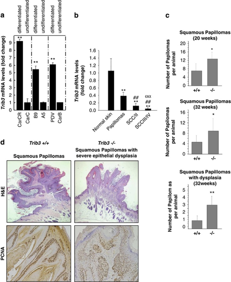
Figure 3.
TRIB3 genetic inactivation accelerates the malignant progression of mouse skin papillomas. (a) TRIB3 mRNA levels as determined by real-time quantitative PCR in different mouse skin cancer cell lines. Data are expressed as the mean fold-increase±S.D. relative to the corresponding less differentiated cell lines (n=3; **P<0.01 compared with undifferentiated cells). (b) TRIB3 mRNA levels as determined by real-time quantitative PCR in samples derived from normal skin, papillomas, squamous cell carcinomas (SCC) grades I and II (SCCI/II) and SCC grades III and IV (SCCIII/IV) obtained from mice subjected to the two-stage model of skin carcinogenesis. Data correspond to TRIB3 mRNA levels and are expressed as the mean fold change±S.D. relative to normal skin (n=8, 8, 6 and 6 for normal skin, papillomas, SCCI/II and SCCIII/IV, respectively; **P<0.01 from normal skin; ##P<0.01 from papillomas and ααP<0.01 from SCC I/II samples). (c) Effect of TRIB3 genetic inactivation on the degree of differentiation and aggressiveness of the skin lesions appeared in Trib3−/− and Trib3+/+ mice subjected to the two-stage skin carcinogenesis protocol (Animals were killed 20 or 32 weeks after the administration of the DMBA treatment and subsequently subjected to histopathological analyses). Data are expressed as the mean number of squamous papillomas or squamous papillomas with dysplasia per animal±S.E.M. (week 20: six Trib3+/+ and five Trib3−/−mice; week 32: seven Trib3+/+ and nine Trib3−/− mice; **P<0.01 or *P<0.05 from Trib3+/+ animals). (d) Representative images of hematoxilin/eosin (upper panel, × 2) and PCNA staining (lower panel, × 10) of squamous papillomas and squamous papillomas with severe dysplasia observed in Trib3+/+ and Trib3−/− mice. See also Supplementary Fig S3

