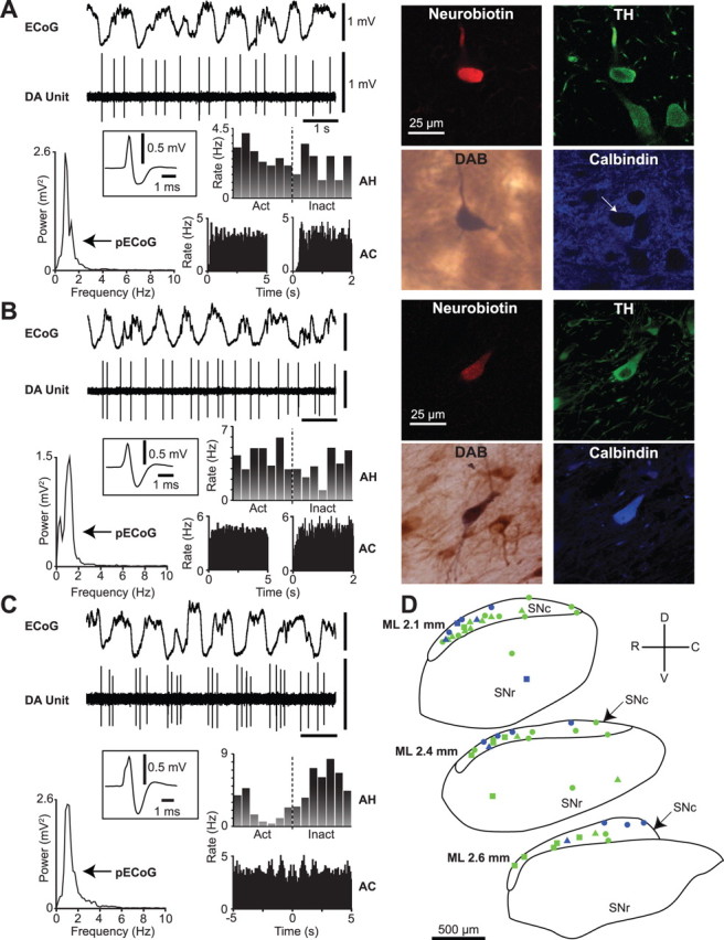
Figure 1.

Spontaneous activity of identified dopaminergic neurons in the substantia nigra. A, Recording of spontaneous unit activity (DA Unit) of a typical calbindin-negative dopaminergic neuron during robust slow-wave activity recorded in the ECoG. Cortical activity is dominated by a ∼1 Hz oscillation, as shown in the ECoG power spectrum (pECoG). Unit activity is irregular, as shown by a relatively flat autocorrelogram (AC; left in 50 ms bins, right in 5 ms bins), and the neuron did not fire preferentially during either the active (Act) or inactive (Inact) component of the cortical slow oscillation (activity histogram, AH). Average action potential waveform (filter settings 300–5000 Hz) of the recorded neuron is shown in the inset. After recording, this neuron was revealed to be a calbindin-negative dopaminergic neuron, as shown in the digital micrographs of Neurobiotin, tyrosine hydroxylase (TH), and calbindin reactivity (arrow points to neuron). Neurobiotin was later visualized with diaminobenzidine (DAB) to form a permanent reaction product for light microscopy. B, A typical calbindin-positive dopaminergic neuron. Note the similarity in electrophysiological characteristics between units in A and B. C, An identified calbindin-negative dopaminergic neuron that showed oscillatory activity at ∼1 Hz, as shown by the peaks in the ACs. This neuron preferentially fired in time with the inactive component of the cortical slow oscillation, as shown in the AH. D, Schematic of sagittal sections of the substantia nigra pars compacta (SNc) and substantia nigra pars reticulata (SNr) showing approximate positions of all identified calbindin-negative (green, n = 40) and calbindin-positive (blue, n = 16) dopaminergic neurons, and whether their responses to a pinch stimulus (circles) or pinch and electrical stimuli (triangles) were tested or not (squares). Note that the calbindin-positive neurons were located mainly within the dorsal tier of SNc. Calibration of ECoG and DA unit in A also applies to B and C. ML (mediolateral) numbers in C denote positions with respect to midline. R, Rostral; C, caudal; D, dorsal; V, ventral.
