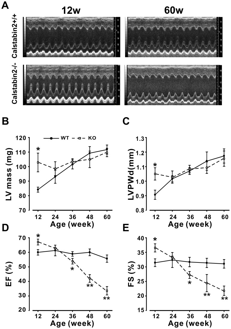
Figure 1. Calstabin2 KO mice exhibit age-dependent heart dysfunction.
(A), Representative echocardiographic (M-mode) photographs from 12- and 60- week-old mice. (B), Echocardiographic measurement of the left ventricle mass (LV mass) at 12, 24, 36, 48 and 60–week-old Calstabin2 KO and WT littermates. LV mass was 22% higher in 12w KO mice than in WT mice, but the aged KO mice displayed similar LV mass, compared to the WT littermates. (C), Ultrasound assessment of left ventricular posterior wall at diastole (LVPWd) in KO and WT mice. (D), Echocardiographic analyses of the ejection fraction (EF). Notably, EF was greatly elevated at the age of 12 weeks, but decreased at 36, 48 and 60 weeks compared to WT littermates. (E), Echocardiographic evaluation of fractional shortening (FS) in 12, 24, 36, 48 and 60–week-old KO and WT littermates. Data are presented as the means ± s.e.m.; n = 6 to 8 per group; *p < 0.05, **p < 0.01.

