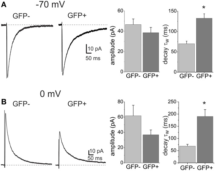
Figure 1.
GABAA receptor-mediated eIPSCs differ in their decay kinetics between lamina II dorsal horn neuron subpopulations. (A) IPSCs evoked by focal stimulation recorded from a EGFP negative (GFP−) and a EGFP positive (GFP+) neuron. Recordings were made at a −70 mV holding potential with CsCl containing pipettes. Histograms show the mean amplitude and weighted decay time constant (τw) in the two cell populations. The asterisk denotes significant difference (n = 21 for GFP− and n = 28 for GFP+). (B) Example traces of eIPSCs recorded from GFP− and GFP+ cells at a holding potential of 0 mV and using CsSO3CH3-containing patch pipettes. Histograms show the mean amplitude and decay τw. Asterisk denotes significant difference (n = 12 for GFP− and n = 14 for GFP+).

