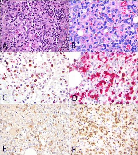
Figure 1.

Morphology and phenotype of TNKLPD. Case with polymorphic infiltrate composed of mixed population of small lymphoid cells with minimal atypia, histiocytes and eosinophils (A, H&E, original magnification 600×). Monomorphic infiltrate consisting of large malignant lymphoid cells with irregular nuclei (B, H&E, original magnification 600×). Expression of cytotoxic marker TIA1 in EBER-positive tumour cells (C, EBER/TIA1 double stain, EBER stains nucleus brown and TIA1 stains cytoplasmic granules red, original magnification 1000×). Positive expression for EBER in situ hybridization in CD3-positive tumor cells (D, EBER/CD3 double stain, EBER stains nucleus brown and CD3 stains cell membrane/cytoplasm red, original magnification 1000×). Positive expression for CD56 (E, CD56, original magnification 600×) and TCRbeta (F, TCRB, original magnification 600×). All photographs were taken with a DP20 Olympus camera (Olympus, Tokyo, Japan) using an Olympus BX41 microscope (Olympus). Images were acquired using DP Controller 2002 (Olympus) and processed using Adobe Photoshop version 5.5 (Adobe Systems, San Jose, CA, USA).
