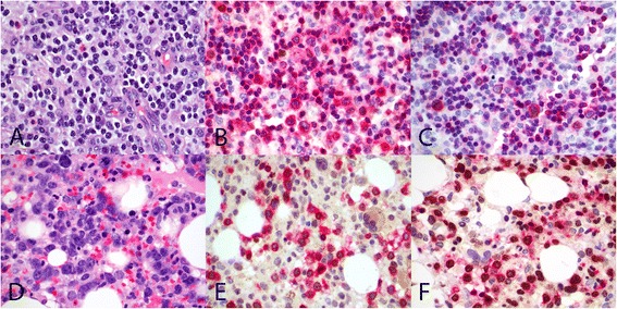
Figure 5.

Immunohistochemical expression of cyclin E2 protein and Ki67 proliferation rate in M-group compared to P-group cases. Case 7 with type A2 disease and polymorphic morphology (P-group) (A, H&E original magnification 600×) showing low cyclin E2 expression (B, CD3/cyclin E2 double stain, original magnification 600×) and low Ki67 proliferation (C, CD3/Ki67 double stain, original magnification 600×). Case 19 with type B disease and monomorphic morphology (M-group) (D, H&E original magnification 600×) with moderately high cyclin E2 expression (E, CD3/cyclin E2 double stain, original magnification 600×) and high Ki67 proliferation (F, CD3/Ki67 double stain). CD3 stains cell membrane/cytoplasm red, and cyclin E2 and Ki67 stain nucleus brown. All photographs were taken with a DP20 Olympus camera (Olympus, Tokyo, Japan) using an Olympus BX41 microscope (Olympus). Images were acquired using DP Controller 2002 (Olympus) and processed using Adobe Photoshop version 5.5 (Adobe Systems, San Jose, CA, USA).
