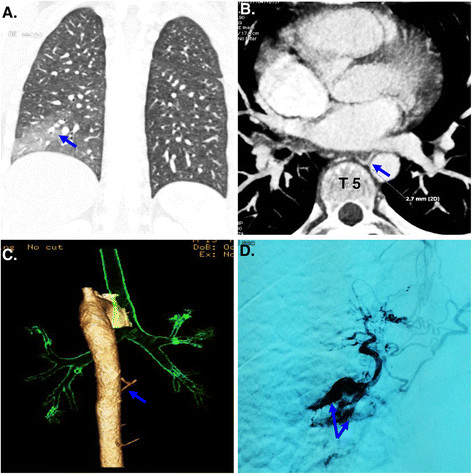
Figure 1.

Radiolographic findings of Dieulafoy’s disease of the bronchus. (A) A chest CT showed exudative lesion, consolidation and atelectasis of the right lower lobe; (B, C) A CT angiography of bronchial artery showed a right bronchial artery arising from the anterior wall of the thoracic aorta at T5 level; (D) Angiography of the bronchial artery showed that a distal right branch of the bronchial artery arising from the thoracic aorta at T5 level was dilated and tortuous.
