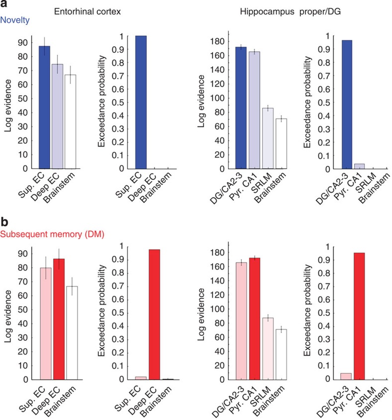
Figure 3. Bayesian model comparisons for decoding of novelty and subsequent memory in entorhinal and hippocampal layers/subregions.
MVB decoding was used to test whether novelty or subsequent memory (‘DM’) can be better predicted from activity in one region than another. The method allows use of the individual (not normalized, not smoothed) functional data to compute the log evidences for each subject-specific anatomical mask (N=20). The relative probability of one model (subregion) over any other model tested is expressed in terms of the exceedance probability (xp) based on random-effects BMS. As control region, the anterior brainstem was included in all analyses. (a) Novelty: Novel trials were detected with highest probability from activity in superficial (‘input’) subregions within the EC (xp: superficial EC=100.0%, deep EC=0%, brainstem=0%) and from DG/CA2–3 within the hippocampus proper/DG (xp: DG/CA2–3=96.3%, pyr. CA1=3.7, SRLM=0.0%, brainstem =0). (b) DM: In contrast, subsequent memory was better decoded from deep (‘output’) subregions within the EC (xp: superficial EC=2.1%, deep EC=97.8%, brainstem=0.1%) and pyramidal CA1 (‘output’) layers within HC proper/DG (xp: DG/CA2–3=4.8%, pyr. CA1=95.2, SRLM=0.0%, brainstem =0.0). Please note that hippocampal analyses were restricted to the hippocampal body where subfields and CA1 apical (‘SRLM’) and pyramidal layers could be reliably differentiated. Error bars denote s.e.m.

