Figure 6.
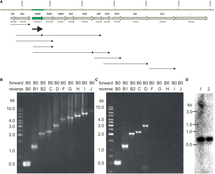
Transcripts observed for the P. furiosus flagellar operon. (A) Flagellar operon of Pyrococcus furiosus with neighboring genes in the upper part. All genes are transcribed from the negatively oriented DNA strand, but are shown here from left to right for easier orientation. Arrows in the lower part indicate cotranscripts identified via RT-PCR. (B) PCR data using genomic DNA as positive control; forward primer was Pfu-flaB0_f, reverse primers were Pfu-flaB0_r to Pfu-flaJ_r. (C) PCR data using cDNA after reverse transcription of isolated RNA and the same primers as given in (B); (data for all other transcripts are found in Supplementary Figure S1). (D) Northern blot experiments using a flaB0 probe. RNA was isolated from late exponentially growing cells (lane 1) and cells in stationary phase (lane 2) and separated in a denaturing agarose gel. The gel migration behavior of an RNA standard is indicated to the left.

