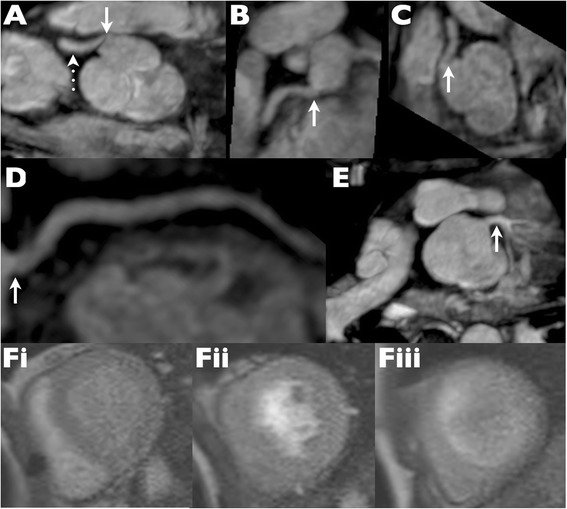
Figure 4.

Evaluation of a “kinked” right coronary artery. (A) Straight axial image from a whole heart magnetic resonance angiogram demonstrates an apparent “kink” at the right coronary artery origin. (B-D) Subsequent multiplanar and centre line reformats however demonstrate that the ostium is in reality unobstructed. (E) The left coronary origin is also unobstructed. (Fi-iii) Stress perfusion magnetic resonance frames at basal, mid and apical left ventricular level show no evidence of any inducible perfusion defect. Quantitative perfusion measured in the right coronary territory was normal (not shown).
