
Figure 2.
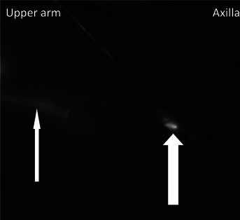
Indocyanine green fluorescence image during the sentinel node biopsy procedure (right axilla). Fluorescence image of the subcutaneous lymphatic channel through the skin (narrow white arrow). Axillary lymphatic flow into the breast sentinel lymph node (corresponding) group (thick white arrow).