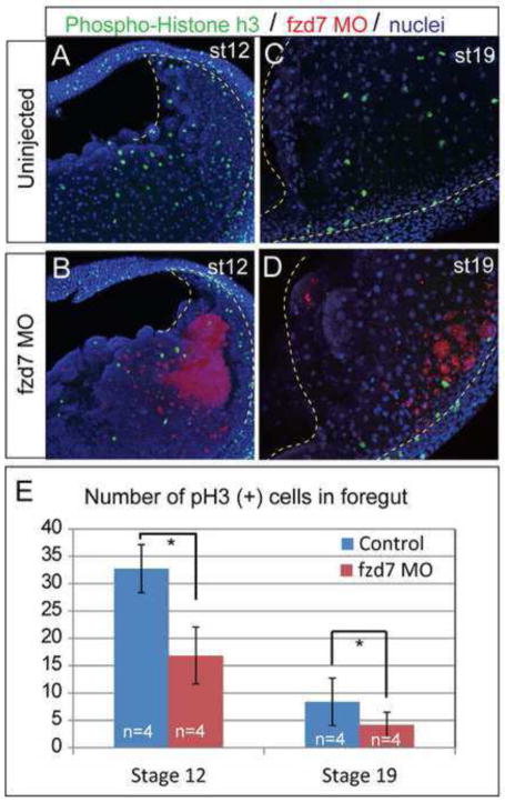
Figure 4. Fzd7-depletion causes reduced cell proliferation.
(A–D) Confocal immunostaining of phospho-Histone h3 (PH3; green), nuclei (blue) and fzd7-MO/RLDx (red) at stage 12 (A,B) and stage 19 (C,D) show that Fzd7-depleted embryos exhibit reduced foregut (outlined in dashed yellow line) proliferation. (E) Mean number of PH3 positive cells in the foregut +/− S.D. *p<0.05 relative to sibling controls in Student’s t-test (n= 4 embryos/condition).

