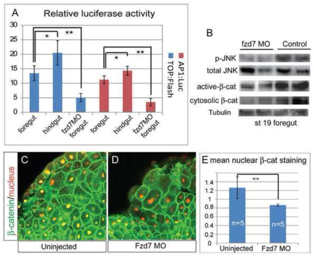
Figure 5. Fzd7 depletion results loss of both Wnt/β-catenin and Wnt/JNK activity in the foregut.
(A) Fzd7-depletion resulted in a reduction of β-catenin/Tcf and JNK/AP1 activity in the foregut. TOP:flash or AP1:Luciferase reporter plasmids were injected into either the D1 foregut endoderm cells or the D4 hindgut endoderm cells at the 32-cell stage, with or without fzd7-MO as indicated. The TOP:Flash reporter is an indicator of β-catenin/Tcf activity, while the AP1:luciferase reporter is an indicator of JNK-mediated c-Jun/c-Fos (AP1) activity. At stage 20 luciferase activity was measured, in triplicate. The average relative luciferase activity, normalized to co-injected pRTK:Renila, from three biological replicates per condition is shown +/− S.D. *p<0.05 and ** p<0.01 relative to control foregut in Student’s t-test. (B) Western blot showed decreased phospho-JNK1/2 (p-JNK) and a loss of dephosphorylated active β-catenin and total cytosolic β-catenin in the foregut explants at stage 19. (C–D): Confocal immunostaining showed reduced nuclear β-catenin levels in Fzd7 morphant foregut tissue (D) relative to controls (C), at stage 20. (E) Mean pixel intensity of nuclear β-catenin staining measured using Image-J +/− S.D (foregut cells were scored from 5 embryos/condition).

