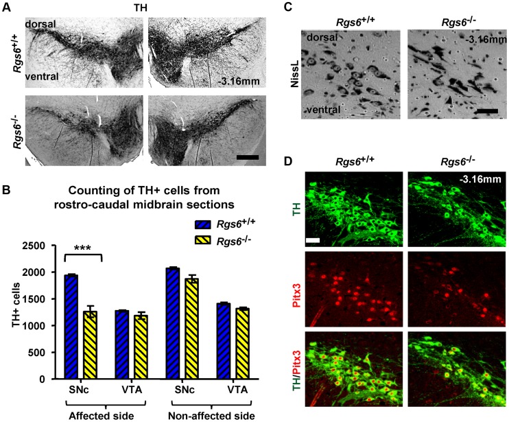
Figure 4. Unilateral loss of Pitx3-positive dopaminergic neurons in ventral SNc of Rgs6 −/− mice.
(A) Immunoperoxidase staining for TH on representative coronal midbrain sections showing less SNc TH+ neurons on one side of Rgs6 −/− mice at 1 year of age compared to sib control. Sections are identified with Bregma position. Scale bar 400 µm. (B) Number of TH+ cells in SNc and VTA of TH-stained coronal sections across midbrain (every 30 µm). Cell counts are represented as means +/− S.D. (***p<0.005). (C) Nissl staining of vSNc sections contiguous to A shows fewer cell bodies and abnormal elongated neurons in Rgs6 −/− mice compared to control. Scale bar 100 µm. (D) Double immunofluorescence staining for TH (green) and Pitx3 (red) on sections contiguous to A showing a marked loss of TH+Pitx3+ cells in Rgs6−/− mice. Scale bar 100 µm.

