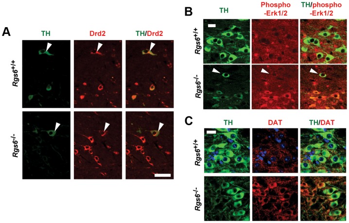
Figure 8. Drd2-related changes of gene expression in degenerating neurons of vSNc.
(A) Immunohistofluorescence staining for TH and Drd2 in coronal sections of 1 y-old Rgs6−/− mice and WT controls. mDA neurons of vSNc express higher levels of glycosylated dopamine receptor D2 (Drd2) than those of dSNc (arrowheads). Scale bar 50 µm. (B) Phospho-Erk1/2 (red) staining is only present in degenerating vSNc THlow (green) neurons of Rgs6−/− mice and not in WT controls. Scale bar 20 µm. Arrowheads indicate unaffected neurons. (C) DAT (red) staining is stronger in degenerating vSNc THlow (green) neurons of Rgs6−/− mice than in WT controls. Scale bar 20 µm.

