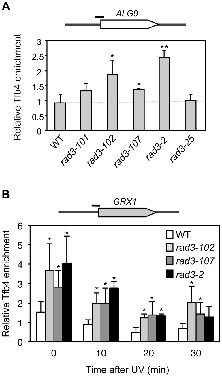
Figure 3. Analysis of TFIIH recruitment to promoters in rad3 mutants.
(A) Chromatin Immunoprecipitation (ChIP) analysis of Tfb4-TAP. Cells were grown in synthetic complete (SC) medium until the exponential phase. ChIP analysis was performed at the ALG9 promoter and normalized with respect to the MFA2 promoter in MAT α cells, which is constitutively repressed. (B) ChIP analysis of Tfb4-TAP after UV damage. Cells were grown in SC medium until the exponential phase, and then irradiated with 80 J/m2. Analysis of the different time-point samples was performed at the GRX1 promoter and normalized with respect to the MFA2 promoter in MAT α cells, which is constitutively repressed. The mean and the SD of triplicate assays of four independent experiments are depicted for each condition. *, p<0.05, **, p<0.01 (Student's t-test).

