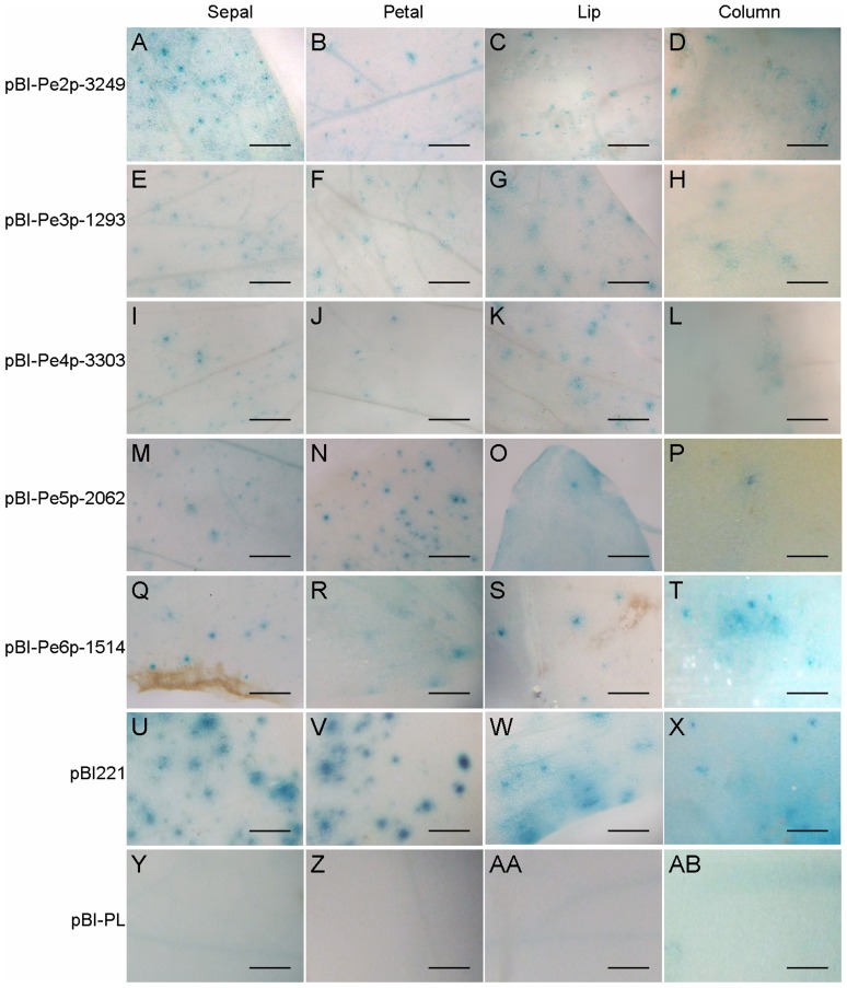
Figure 3. GUS histochemical staining for the promoter activities of PeMADS2∼6.
Histochemical assay of GUS expression in floral organs shown in the order of pBI-Pe2p-3249 (A–D), pBI-Pe3p-1293 (E–H), pBI-Pe4p-3303 (I–L), pBI-Pe5p-2062 (M–P), pBI-Pe6p-1514 (Q–T), pBI221 (U–X) and pBI-PL (Y-AB). Constructs were bombarded into four independent floral buds, and results are representative of three independent bombardment experiments. Scale bar = 0.5 mm.

