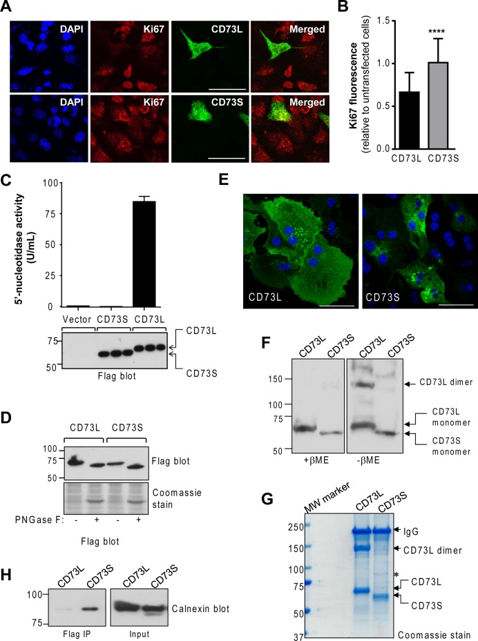
FIGURE 5:
Molecular and functional characteristics of CD73S. (A) Coimmunofluorescence analysis of the proliferation marker Ki67 (red) and Flag-CD73S/L (green) in transfected HepG2 cells. Blue, 4′,6-diamidino-2-phenylindole (DAPI); bar, 20 μm. (B) Quantification of Ki67 immunofluorescence staining in CD73-expressing cells relative to neighbor untransfected cells (30 CD73+ cells and 150 CD73− untransfected cells were counted per condition). ****p < 0.0001, unpaired t test. (C) Measurement of 5′-nucleotidase activity in transfected HEK293T cell lysates. Samples expressing equal protein levels (bottom) were analyzed in triplicate. (D) Treatment of CD73-transfected HEK293T cell lysates with peptide-N-glycosidase F results in similar deglycosylation of both isoforms. (E) Immunofluorescence-based localization of Flag-CD73L or Flag-CD73S in primary mouse hepatocytes. Blue, DAPI; bar, 20 μm. (F) Flag immunoblot of Flag-CD73L– and -CD73S–expressing HEK293T total cell lysates analyzed under reducing (+β-mercaptoethanol, βME) or nonreducing (–βME) conditions, showing that CD73S does not dimerize. (G) Coomassie-stained gel of Flag immunoprecipitates of the nonreduced samples shown in F. Asterisk denotes a unique protein present in CD73S immunoprecipitates, identified by mass spectrometry as calnexin. (H) Biochemical validation that CD73S coimmunoprecipitates with calnexin.

