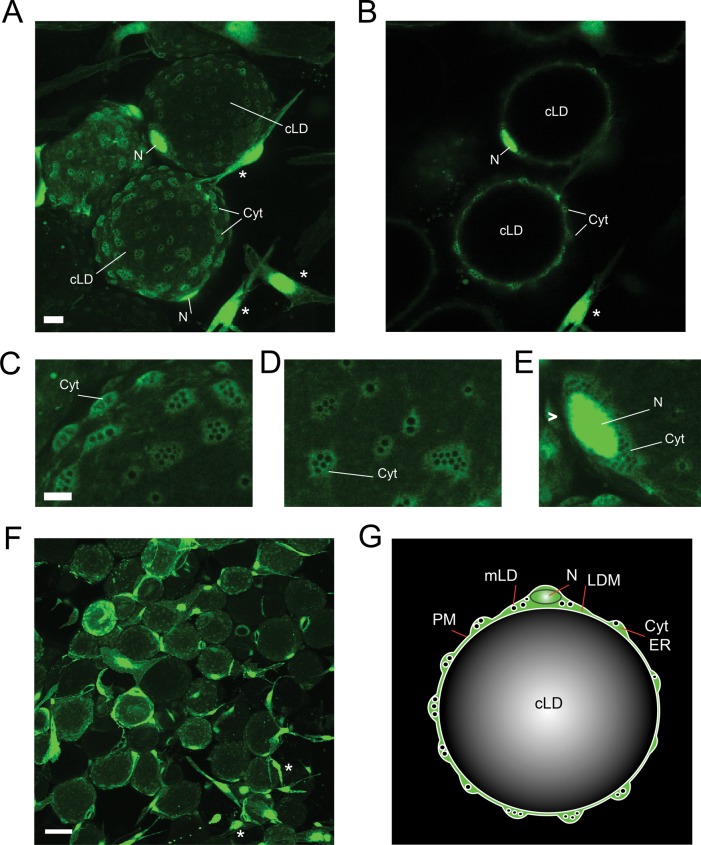
FIGURE 1:
Cytoarchitecture of primary unilocular adipocytes. Visceral WAT explants were infected with an adenoviral vector encoding eGFP as described in Materials and Methods. On day 2 after infection, live explants were examined for GFP expression using confocal microscopy. cLD, central LD; Cyt, cytoplasm; LDM, lipid droplet membrane; mLD, micro-LD; N, nucleus; PM, plasma membrane. (A) A field of view shows GFP-positive unilocular adipocytes (spheres) and stromovascular cells (asterisks) residing in WAT. The image represents the sum of confocal slices. Bar, 10 μm. (B) Single confocal section of the image in A. Enlarged areas of adipocytes containing cytoplasmic nodules (C, D) and perinuclear cytoplasm (E) filled with GFP-negative spherical cavities. Bar, 5 μm. (F) Portion of the GFP-infected WAT explant shown at lower magnification. Bar, 50 μm. (G) Schematic representation of the spatial organization of a unilocular adipocyte. The cLD of a unilocular adipocyte represents a single sphere tightly fitted inside the cell, whereas the cytoplasm forms multiple nodules containing various organelles.

