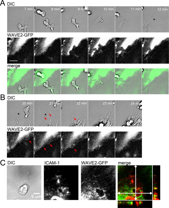
FIGURE 2:
WAVE2 localization to membrane protrusions that close gaps and form at sites of transcellular pores. (A) WAVE2 is not recruited to paracellular pores during PBL transmigration. Frames from Supplemental Movie S2. Asterisk indicates a gap between ECs that persists after transmigration. Scale bar, 3 μm. (B) WAVE2 localizes to endothelial monolayer gaps that close after lymphocyte transmigration. Frames from Supplemental Movie S3. Asterisks indicate the gap, which closes after a wave of membrane protrusion. Red arrows indicate membrane waves, where WAVE2 has accumulated. Scale bar, 3 μm. (C) WAVE2 localizes to docking structures at sites of transcellular migration. ECs expressing WAVE2-GFP and immunolabeled for ICAM-1. Transmigrating lymphocyte is shown in the DIC image. The yz- and xz-projections from a 3D data set reveal WAVE2 in membrane protrusions that cup the PBL.

