FIGURE 1:
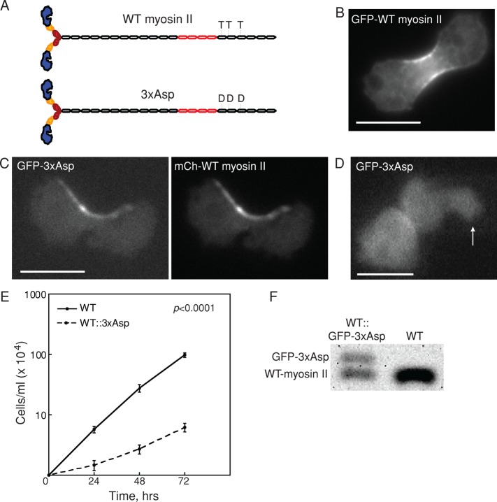
Overexpression of 3xAsp in WT cells reduced growth rate. (A) Diagram of WT myosin II and 3xAsp myosin II. (B) GFP-WT myosin II accumulates at the furrow. Scale bar, 10 μm. (C) Myosin II distribution in myoII::GFP-3xAsp;mCh-WT myosin II was altered by expression of 3xAsp myosin II. GFP-3xAsp and mCherry-WT myosin II colocalized at the cleavage furrow in dividing cells but with an aberrant distribution. Scale bar, 10 μm. (D) WT cells expressing GFP-3xAsp (WT::3xAsp) did not show mechanosensitive myosin II accumulation in response to micropipette aspiration (white arrow). The background-corrected fluorescence intensity ratio of the cortex inside the micropipette (Ip) and the opposite cortex (Io) was measured and used to determine whether the myosin II undergoes mechanosensitive accumulation (Effler et al., 2006). Scale bar, 10 μm. (E) Suspension culture growth for WT control and WT::3xAsp cells. Average growth rate is 0.067 ± 0.008 h−1 for WT cells (n = 11) and 0.032 ± 0.009 h−1 for WT::3xAsp cells (n = 11; p value is on the graph). (F) Western analysis of WT and WT::GFP-3xAsp (where 3xAsp is integrated randomly in the genome) showed that 3xAsp is 40% and WT endogenous myosin II is 60% of the total myosin II in these cells.

