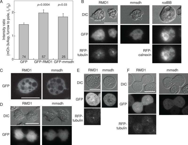
FIGURE 4:
Localization of RMD1, mmsdh, and rcdBB proteins in cells. (A) Bar graph shows the cleavage furrow intensity ratio of mCherry-3xAsp myosin II in myoII::mCh-3xAsp cells when GFP-RMD1 and GFP-mmsdh were expressed. These data confirm that GFP-RMD1 and GFP-mmsdh are functional GFP-fusion proteins. Sample sizes and p values are displayed on the graph. (B) Epifluorescence images demonstrate subcellular localization of RMD1, mmsdh, and rcdBB. RMD1 is largely cytoplasmic, with some enrichment around the centrosome (RFP-tubulin is shown for comparison). mmsdh is only cytosolic, and rcdBB was found enriched in the endoplasmic reticulum (RFP-calnexin is shown for comparison). (C) Confocal imaging confirms the largely cytosolic distribution of RMD1, with weak enrichment around the centrosome and the cytosolic distribution of mmsdh. (D) During cytokinesis, RMD1 and mmsdh remain cytosolic, with RMD1 showing weak enrichment around the centrosome. (E) In interphase cells compressed by agarose overlay, which introduces mechanical stress to the cortex, RMD1 remains largely cytosolic, with weak enrichment around the centrosome (RFP-tubulin is shown for comparison). mmsdh also remains cytosolic. (F) In dividing cells compressed by agar overlay, RMD1 and mmsdh remain cytosolic, with RMD1 showing weak enrichment around the centrosome (RFP-tubulin is shown for comparison).

