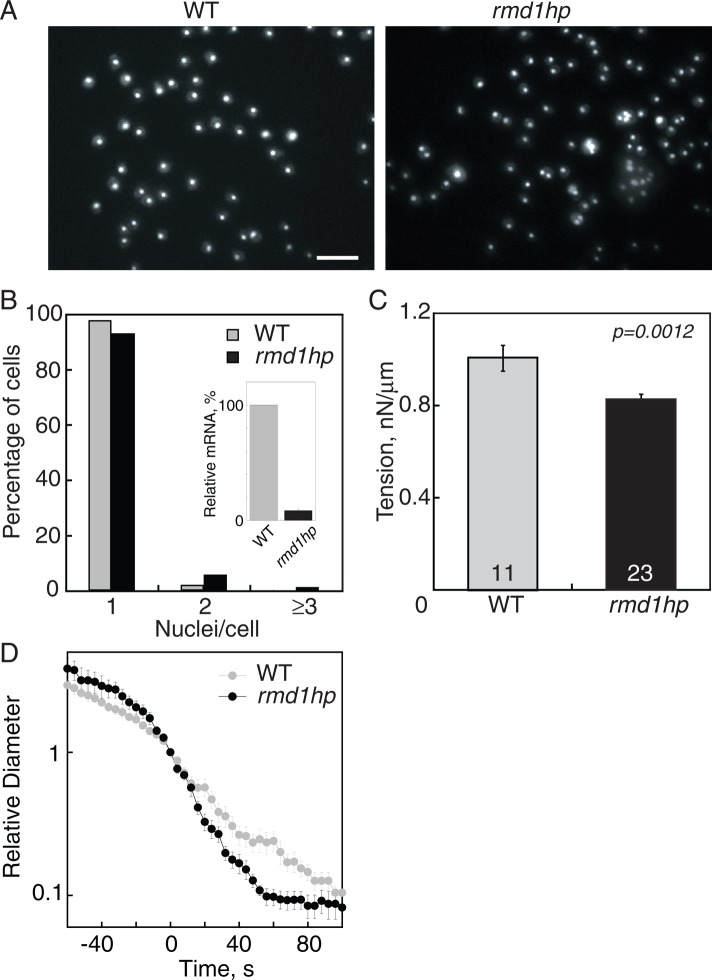
FIGURE 5:
Depletion of rmd1 mRNA led to cytokinesis and cortical tension defects. (A) Micrographs of 4′,6-diamidino-2-phenylindole (DAPI)-stained cells show that rmd1hp cells have more multinucleated cells than WT control cells. Scale bar, 50 μm. (B) Frequency histogram reveals an increase in the fraction of multinucleated cells in rmd1hp cells. Inset demonstrates the 91% depletion of rmd1 mRNA in rmd1hp cells. (C) The rmd1hp cells had a 20% reduction in cortical tension. (D) Semilog plot of the furrow ingression dynamics of WT vs. rmd1hp cells, showing that the rmd1hp cells had a faster, more linear furrow ingression dynamic than WT control (n = 8–10 cells/genotype).

