FIGURE 1:
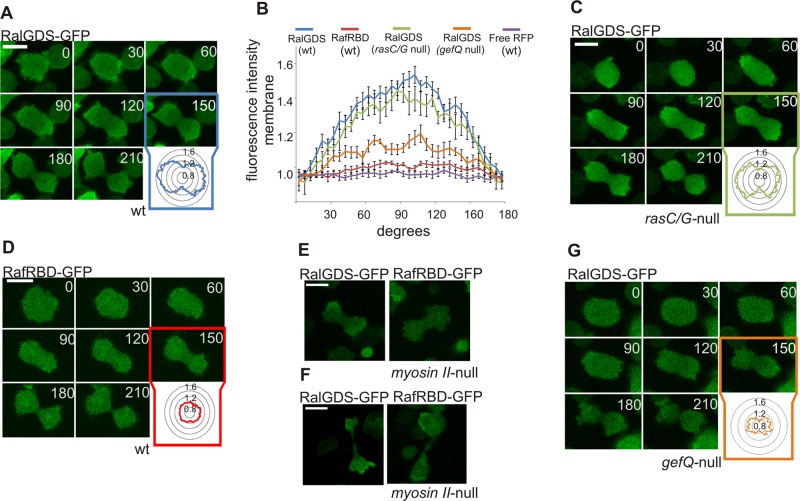
Dynamic Rap1 activation during cytokinesis. (A) Images of RalGDS-GFP (detecting active Rap) in dividing Dictyostelium wild-type cells. Inset: RalGDS-GFP fluorescence intensity was measured at the cell boundary around the circumference of the cell relative to the fluorescence intensity in the cytosol. (B) Quantification of the average fluorescence intensities of the indicated fluorescent markers along the cell membrane of dividing Dictyostelium cells presented as degrees from the cleavage furrow (see A, C, D, and G for representative images of the experiments). Error bars represent SEM. (C) Image of RalGDS-GFP in rasC/rasG-null cells. (D) Images of RafRBD-GFP (active Ras) in dividing Dictyostelium wild-type cells. Images of RalGDS-GFP or RafRBD-GFP in myosin II-null cells dividing by type B (E) and type C (F). (G) Image of RalGDS-GFP marker in gefQ-null cells. Scale bars: 10 μm.

